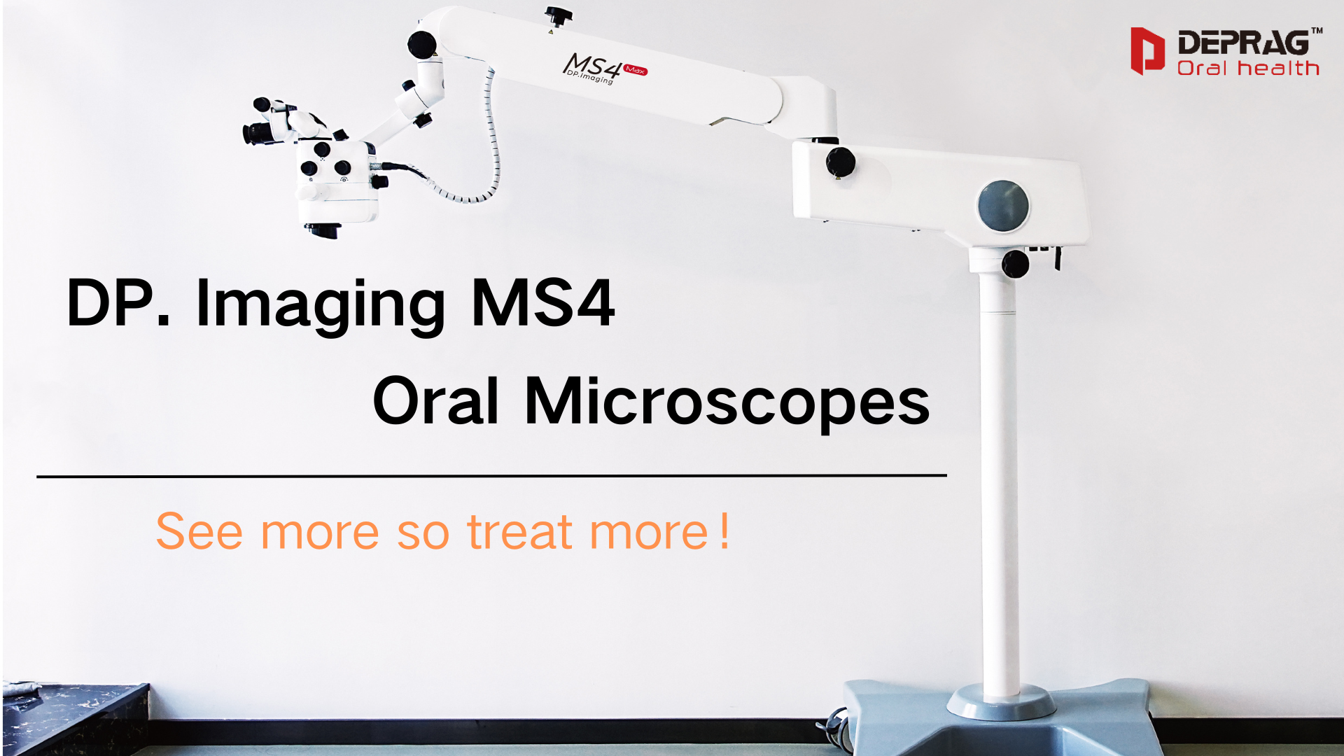
A refined treatment solution specially developed for dental clinical diagnosis and treatment.

With the development of the medical field, the original “one-size-fits-all” diagnosis and treatment method cannot meet the needs of patients. Instead, a series of specific restorative therapies are used to perform precise cutting operations on dental tissue. However, the naked eye cannot meet the needs of the operation, so Dental clinical equipment with magnification and lighting functions emerged at the historic moment, and the oral microscope is a product of development and improvement.
Application Of Oral Microscopy
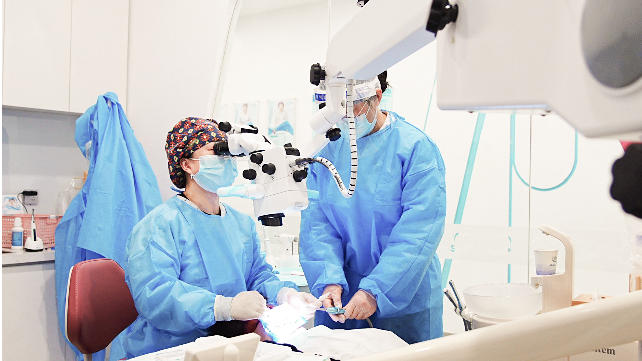
Oral microscopes are widely used because they can magnify at higher magnifications, provide more details, and enhance the visualization of surgical operations. Dental doctors can operate more accurately and optimize the operation process.Oral microscopes are currently used in:
- Root Canal Therapy
- Tooth Decay
- Dental lmplant
- Veneers
- Deep Cleaning

Support System For Oral Microscopy
1.Lighting system
The bright lighting environment provides doctors with a clearer vision and relieves eye fatigue, and different filter lenses meet the needs of different dental departments.
2.Stent system
The adjustable stent can not only make reasonable use of space in the outpatient end, facilitate transportation and storage, but also adjust the position according to the individual differences of doctors, which is in line with human engineering and reduces occupational hazards.
3.Digital imaging system
Real-time reflection of the internal images of the oral cavity makes communication between doctors and patients less difficult and more intuitive. The storage function facilitates case archiving and later learning and communication.
4.Amplify system
The wide range of adjustable focus provides doctors with higher-definition detailed images to help perform precise operations.
Benefits Of Using An Oral Microscope
Oral microscopes support doctors in the clinical field, protecting tooth structure, preserving tissue, minimizing risks and shortening patient recovery times through ultra-magnified images.

- The high magnification view has excellent and realistic visual effects, and the precision and accuracy of operation are greatly improved.
- The design of ergonomics is to reduce the risk of occupational diseases and improve the work efficiency of doctors.
- Maintain a comfortable working distance with patients to improve patients’ medical experience.
- Real-time imaging enables patients to intuitively understand their oral conditions and reduces the communication costs of doctors.
- Built-in high-definition image and video recording function, help later training and communication.
- The treatment results of patients were recorded in stages and stored in files, so that the treatment effect could be clearly recorded.
Why Choose DP.Imaging MS4 Oral Microscope?
DEPRAG Oral Health is committed to helping the fine development of oral clinical field, achieving higher accuracy in each link, so that patients have a better treatment experience.
DP.Imaging MS4 is jointly developed and produced by China National Academy of Optical Systems, and can be widely used in the field of general dentistry. This microscope oral microscope features world-class accessory elements that help clinicians more clearly observe the fine structure of the oral system and add ergonomic design to improve clinical work efficiency, significantly improve operational processes, and enable physicians to achieve a more comfortable work throughout their careers.